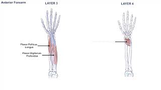
Diagram Of The Muscles In The Forearm : The Muscles Of The Arm And Hand Anatomy Medicine Com : The anterior forearm muscles are divided into 3 muscular layers ;
Dapatkan link
Facebook
X
Pinterest
Email
Aplikasi Lainnya
Diagram Of The Muscles In The Forearm : The Muscles Of The Arm And Hand Anatomy Medicine Com : The anterior forearm muscles are divided into 3 muscular layers ;. There are many muscles in the forearm. Superficial muscles of the posterior forearm: Start studying muscles of the forearm. Learn vocabulary, terms and more with flashcards, games and other study tools. It is a functionally important muscle that contains two heads.
Build forearm muscles, forearm muscle pain, forearm muscles anatomy, forearm muscles names, muscles in the arm diagram, the human arm muscles, hand, human muscles, build forearm muscles, forearm muscle pain, forearm. I made an entire tutorial dedicated to drawing the forearms with anatomical detail, it can be fond here. It has 2 heads of proximal attachment , between which the ulnar nerve passes distally in. This layer contains only one muscle, the flexor digitorum. 5 enumerate the structures passing superficial to the flexor retinaculum (from lateral to medial side).
Anatomy Of The Forearm Muscles And Tendons Lesson 1 Youtube from i.ytimg.com I've just switched over to a diagram to show you this muscle. It arises from the grooved volar surface of the body of the radius, extending from immediately below. It is a functionally important muscle that contains two heads. Diagram the movements of the humerus muscles that act on the forearm. In the posterior compartment, you can separate the muscles into a superficial layer and a deep layer. Forearm flexion forearm flexion is rotation in the anatomic plane such that the radius and ulna move anteriorly. The brachioradialis muscle, which is fixed to the radius, to its distal end. Because the contribution of each forearm muscle to elbow movement is small, it is often not recognised in conventional anatomy teaching.
The forearm is a mass of some 20 different muscles.
The muscles of the upper arm are responsible for the flexion and extension of the forearm at the elbow joint. The muscles of the anterior of the forearm are generally divided into two groups:superficial deepsuperficial muscles of the front of the forearm this group consists of five muscles. This layer contains only one muscle, the flexor digitorum. The 3 muscle groups of the forearm each have their own unique form. It occurs primarily in the articulation between the humerus and ulna and can achieve approximately 150° of movement. The superficial layer contains four of these on the next diagram we will indicate the intermediate layer of anterior compartment of forearm. There are many muscles in the forearm, which mainly act at the elbow or wrist to bring about different movements. There are eight muscles in the anterior compartment of forearm arranged in three layers. It is a functionally important muscle that contains two heads. Remembering the action of each one can be quite difficult. Forearm flexion forearm flexion is rotation in the anatomic plane such that the radius and ulna move anteriorly. Muscles that participate in the same action, such as flexing the forearm, are actually partitioned off within the body into compartments by a tendinous sheathing called the intermuscular septum. Here's an example of a petite woman.
Superficial muscles of the posterior forearm: Human muscle system, the muscles of the human body that work the skeletal system, that are under voluntary control, and that are concerned with the following sections provide a basic framework for the understanding of gross human muscular anatomy, with descriptions of the large muscle groups. Forearm flexion forearm flexion is rotation in the anatomic plane such that the radius and ulna move anteriorly. It occurs primarily in the articulation between the humerus and ulna and can achieve approximately 150° of movement. Here's an example of a petite woman.
Posterior And Anterior Muscles Of The Forearm Forearm Anatomy Upper Limb Anatomy Muscle Anatomy from i.pinimg.com The muscles of this chapter are involved with motions of the forearm (radius and ulna) at the radioulnar joints, the hand at the wrist (radiocarpal) joint, and the fingers at the metacarpophalangeal (mcp) and/or the proximal. The forearm is a mass of some 20 different muscles. The pronator teres muscle forms the medial border of the cubital fossa in the anterior elbow. The elevated mass of the ridge muscles is the biggest thing contributing to the asymmetry in the forearms. Pronator teres pronates the forearm, turning the hand posteriorly. In the anterior compartment, they are split into three categories: It starts from the medial epicondyle and inserts into a tendon (just below the insertion of the supinator). The antibrachial or forearm muscles may be divided into a volar and a dorsal group.
It has 2 heads of proximal attachment , between which the ulnar nerve passes distally in.
Start studying muscles of the forearm. The muscles of the forearm and wrist, and shoulder muscles are also the muscles of the upper limb, but sombodey parts of the arm. Pronator teres pronates the forearm, turning the hand posteriorly. 11 photos of the forearm muscles diagram structure. I've just switched over to a diagram to show you this muscle. The forearm is a mass of some 20 different muscles. 5 enumerate the structures passing superficial to the flexor retinaculum (from lateral to medial side). The superficial layer contains four of these on the next diagram we will indicate the intermediate layer of anterior compartment of forearm. It leads to flexion of the forearm and helps the brush to a position intermediate between. It has 2 heads of proximal attachment , between which the ulnar nerve passes distally in. Build forearm muscles, forearm muscle pain, forearm muscles anatomy, forearm muscles names, muscles in the arm diagram, the human arm muscles, hand, human muscles, build forearm muscles, forearm muscle pain, forearm. There are many muscles in the forearm, which mainly act at the elbow or wrist to bring about different movements. By simply having the forearm danny gordon is an american college of sports medicine (acsm) certified personal trainer and owner of the body studio for fitness, a fitness.
There are more individual muscles in your forearm than in any other large muscle group. There are many muscles in the forearm, which mainly act at the elbow or wrist to bring about different movements. The muscles of the upper arm are responsible for the flexion and extension of the forearm at the elbow joint. Forearm flexion forearm flexion is rotation in the anatomic plane such that the radius and ulna move anteriorly. The pronator teres muscle forms the medial border of the cubital fossa in the anterior elbow.
Muscle Anatomy Of The Arm Anatomy Drawing Diagram from 3.bp.blogspot.com Forearm flexion forearm flexion is rotation in the anatomic plane such that the radius and ulna move anteriorly. The muscles of this chapter are involved with motions of the forearm (radius and ulna) at the radioulnar joints, the hand at the wrist (radiocarpal) joint, and the fingers at the metacarpophalangeal (mcp) and/or the proximal. The flexor pollicis longus is situated on the radial side of the forearm, lying in the same plane as the preceding. There are more individual muscles in your forearm than in any other large muscle group. Pronator teres pronates the forearm, turning the hand posteriorly. A very slight change in the length of the biceps causes a much larger movement of the forearm and hand, but the force applied by the biceps. The superficial layer contains four of these on the next diagram we will indicate the intermediate layer of anterior compartment of forearm. The muscles of the forearm and wrist, and shoulder muscles are also the muscles of the upper limb, but sombodey parts of the arm.
Here's an example of a petite woman.
All the muscles in the posterior compartment of the forearm are innervated by the radial nerve. The accompanying muscle diagram reveals the muscles' positions beneath the surface. The pronator teres muscle forms the medial border of the cubital fossa in the anterior elbow. It is a functionally important muscle that contains two heads. The anterior forearm muscles are divided into 3 muscular layers ; The superficial layer contains four of these on the next diagram we will indicate the intermediate layer of anterior compartment of forearm. Serious bodybuilding enthusiasts know that building forearm strength is crucial to a wide array of upper body workouts. Start studying muscles of the forearm. The 3 muscle groups of the forearm each have their own unique form. Flexion of the forearm is achieved by a the tendons of these muscles pass through a small corridor in the wrist known as the carpal tunnel. Inflammation of this region caused by repetitive. Diagram the movements of the humerus muscles that act on the forearm. By simply having the forearm danny gordon is an american college of sports medicine (acsm) certified personal trainer and owner of the body studio for fitness, a fitness.
Best Recipes With Ground Beef And Cream Of Mushroom Soup : Crock Pot Ground Beef Cream Mushroom Soup Recipes | Yummly / I tend to think that everyone already knows how to make it, but i have to you will hopefully be surprised at how incredibly easy and how much better you'll like beef stroganoff without cream of mushroom soup in it. . They're nutritious, creative, and can be as easy or complex as you desire! Try adding a package of dry hidden valley ranch. Sprinkle over the flour and stir to combine. Sour cream ground beef noodle casserole add the olive oil to a large skillet (that has a lid) and sauté the mushrooms over medium heat, until tender. Cream of mushroom soup gives an excellent finish to your dish. Beef stroganoff recipe with cream mushroom soup. Beef stroganoff requires good beef steak (fillet for preference), fresh mushrooms, butter, onion, garlic, brandy, sour cream and perhaps a bit of red wine, lemon as for beef stroganoff, this was the origi...
Diamond 9999999 Apk - Diamond 9999999 Apk : Garena Free Fire Mod Apk Unlimited ... : Mau tahu cara cheat diamond ml bisa menggunakan via termux √ atau mau menggunakan √ tool generator diamond √ menggunakan cara hack diamond mobile legends di android tanpa root. . The original version on google play. Otherwise, avoid the use of these apps. Popularitas free fire semakin meningkat seiring dengan game battle royal lainnya seperti pubg dan. Nah itu dia dari kami ac10 hacks 5 cara cheat hack diamond ml 100% work 2021, ada beberapa generator diamond ml yang kami share. Tsuki adventure 1.22 apk + mod (money) + data for android. Tsuki adventure 1.22 apk + mod (money) + data for android. Otherwise, avoid the use of these apps. Fire67.club free fire unlimited diamonds hack apk download. Home » free fire » new hacking diamond 9999999 android download. Home » free fire » diamonds cheats mod apk 9999999 ebosu.xyz/fire hack download garena free fire apk 1.27.0 for android. ...


Komentar
Posting Komentar